

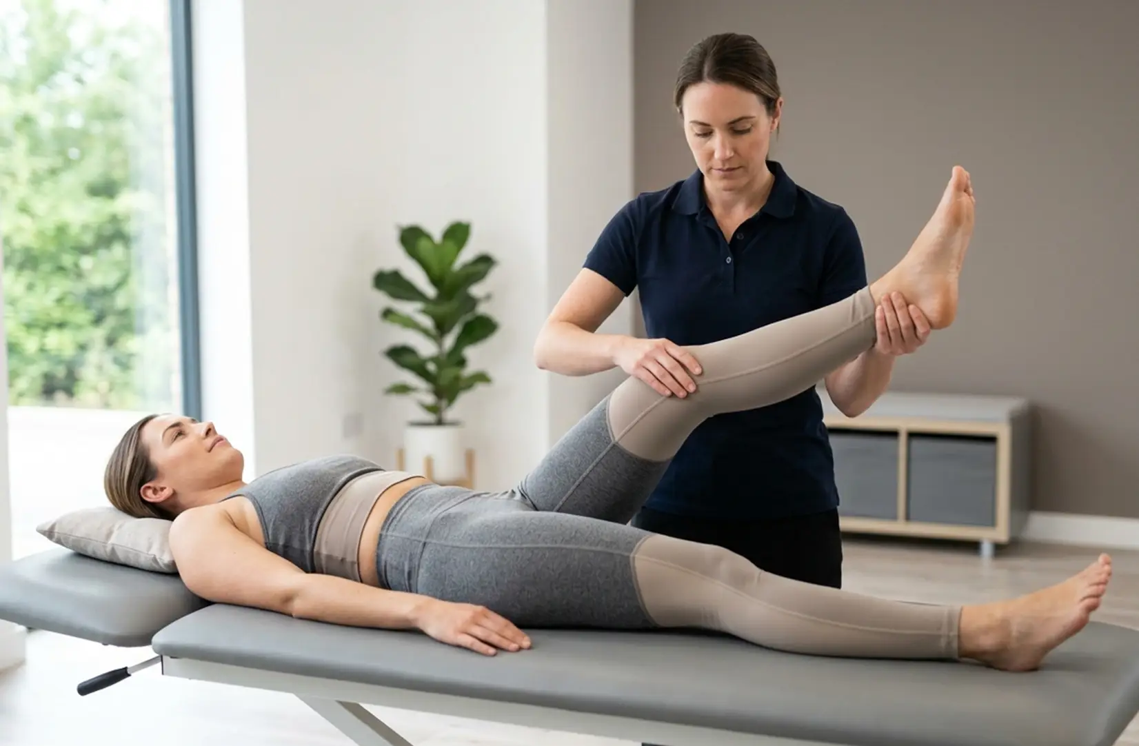
Sciatica Assessment: What a Thorough Examination Actually Involves
Most people who come for a sciatica assessment have already had some form of diagnosis. A GP has told them the nerve is irritated. A scan has shown a disc bulge. Someone has named what they have. What most of them have not had is an explanation of where that diagnosis came from and what it actually means for their recovery.
That gap between label and understanding is where a lot of unnecessary worry lives. People arrive unsure whether movement is safe. They have read conflicting things online. Some have been told to rest. Others have been told to push through. The assessment is where that uncertainty starts to lift.
People are often surprised by how much of the assessment involves conversation rather than tests. The way symptoms behave over a day, which positions ease things, how the problem started and what has changed since then: these details carry more diagnostic weight than any single physical test.
Why Assessment Changes Everything
Sciatica is not a diagnosis in the strict sense. It is a description of symptoms: pain or altered sensation travelling down the leg, usually from the lower back or buttock into the thigh, calf, or foot. The important clinical question is not whether someone has sciatica but what is generating it.
That distinction matters because the source of nerve irritation determines the treatment. A disc prolapse, a narrowed canal compressing the nerve root, a tight piriformis muscle irritating the sciatic nerve in the deep gluteal space, and referred pain from a stiff sacroiliac joint can all produce similar symptoms. Treatment directed at the wrong source often explains why symptoms persist despite otherwise good rehabilitation.
Stynes et al. (BJGP, 2018) found that clinical features, not symptom location alone, are what reliably predict nerve root involvement. Assessment findings are interpreted in the context of the person's work, training, recovery demands, and day-to-day functional goals, because those factors influence how nerve irritation settles and which approach is most appropriate.
What the Assessment Involves
A sciatica assessment at Atherapy follows a structured sequence. Each section informs the next, and the clinical picture emerges progressively rather than from separate, disconnected findings.
The Subjective Examination
The first and often longest part of the assessment is listening. Before any physical examination begins, the clinician needs to understand the full symptom picture: where the pain and any altered sensation are located, how they behave across the day, what provokes and relieves them, and how it has all changed since onset.
This part of the assessment also includes screening questions for cauda equina syndrome.
Beyond the red flag screen, the subjective examination looks at symptom behaviour. Pain that worsens with prolonged sitting and eases with walking suggests something different from pain triggered by standing and relieved by flexion. Pain that moves back toward the spine with certain movements is usually a positive finding. In simple terms, it means certain movements consistently improve symptoms rather than aggravating them. Donelson et al. (Spine, 1997) established this directional preference as a reliable prognostic indicator, because it directly guides the home exercise programme from session one.
The Numeric Pain Rating Scale (NPRS) is recorded at this stage as a baseline for tracking progress. The trend matters more than any single number.
The Physical Examination
The physical examination is structured around answering the questions raised by the subjective history. The process is collaborative rather than something simply done to the patient. Each movement is guided and explained, nothing is forced, and many people find that systematic movement testing clarifies their symptoms rather than provoking anything new.
We assess how symptoms behave under load and movement, not simply where pain is felt. Posterior leg pain can arise from several different mechanisms, and those mechanisms respond to different interventions.
What the examination is trying to identify
- Which structures are most likely generating the symptoms
- Whether a directional movement preference is present
- Whether the sciatic nerve is sensitised or structurally compressed
- Whether the sacroiliac joint or deep gluteal space is involved
- Whether there are neurological findings that require urgent review
What the physical examination tests for
- Movement assessment Which directions provoke or ease symptoms and whether centralisation occurs. Centralisation means symptoms move back toward the spine rather than travelling further down the leg. Where it is present, this guides the direction of early loading in the home exercise programme. Based on Donelson et al. (Spine, 1997).
- Neural tension tests The straight leg raise and slump test assess the mobility and sensitivity of the sciatic nerve. A positive finding does not necessarily mean the nerve is damaged. In most cases it reflects irritation and sensitivity rather than structural injury, and nerve irritation settles with appropriate management.
- Neurological examination Manual muscle testing and sensation testing identify whether there is any motor or sensory deficit consistent with a specific nerve root level. Most people assessed do not show significant weakness or sensory loss, even when symptoms are severe. Where deficits are present, this informs decisions about imaging and specialist referral.
- Sacroiliac joint assessment The provocation cluster described by Laslett et al. (Manual Therapy, 2005) is used where the history suggests possible sacroiliac involvement. At least three positive tests from the cluster are required for a provisional diagnosis of SIJ dysfunction. The sacroiliac joint is a commonly missed source of buttock and leg pain in assessments that focus exclusively on the lumbar spine.
- Deep gluteal assessment Where symptoms do not correspond to a clear dermatomal pattern (that is, a specific nerve root distribution), the deep gluteal space is assessed. Martin et al. (Arthroscopy, 2015) described how the sciatic nerve can be compressed by the piriformis muscle or fibrous bands in this region, producing sciatica-like symptoms without spinal pathology. This is one of the more frequently missed sources of leg pain.
The assessment works best as a collaborative process. The information someone brings about how their pain behaves across the day is at least as important as what I find on examination.
Dr Emma Collier PT, DPT, HCPC
Functional Assessment and Outcome Measures
Following the physical examination, a standardised functional assessment is completed using the Patient Specific Functional Scale (PSFS). The person identifies three activities that their symptoms have made difficult and rates their current ability to perform each one on a scale from zero to ten.
These ratings are revisited at four weeks and at discharge, giving a concrete, trackable measure of progress anchored to what matters in that individual's daily life rather than a generic pain score. Where appropriate, findings are also shared with GPs, consultants, or imaging providers.
What a Thorough Assessment Helps to Rule Out
One of the less visible functions of the assessment is differential diagnosis: identifying what the symptoms are not, as well as what they are. Several conditions produce buttock and leg pain that closely resembles nerve root sciatica.
Sacroiliac joint dysfunction, piriformis syndrome, lumbar facet referral, and hip joint pathology can all do this. The clinical picture from a systematic examination usually allows these to be distinguished without imaging. Where the picture is ambiguous or where neurological signs are present, onward referral or imaging may be recommended.
NICE Guideline NG59 (2016, updated 2020) advises against routine imaging for sciatica but recommends it where neurological deficits are present or the clinical picture does not respond as expected. The assessment helps identify which situation applies.
Worsening weakness in the leg should always be assessed promptly. It should not simply be waited out.
What Most People with Sciatica Can Expect
Patients arriving for assessment are often silently asking questions that have not yet been addressed: Will this settle? Am I permanently damaged? Will I need surgery? Is movement making it worse?
For the majority, the honest answer is reassuring. Chiu et al. (Clinical Rehabilitation, 2015) reported that 90% of disc prolapses show significant spontaneous reabsorption within 12 months. Peul et al. (NEJM, 2007) found that surgery and conservative management produce equivalent outcomes at one to two years, with the main difference being the speed of early symptom relief rather than long-term recovery.
The body knows how to recover. The purpose of the assessment is to identify which approach is most likely to support that process safely and efficiently.
Bilateral leg symptoms always warrant prompt assessment and may require urgent referral regardless of how long the problem has been present.
A working diagnosis is a starting point, not a verdict. If the clinical picture changes or does not respond as expected, the diagnosis is revisited and the plan adjusted accordingly.
GOOD PROGNOSTIC SIGNS
Signs that usually respond well to conservative management
- Symptoms that centralise or ease with specific movements
- Leg pain without significant motor or sensory deficit
- Onset following a period of sustained loading or sedentary posture
- Symptoms that are intermittent or positional rather than constant
- No bladder, bowel, or bilateral involvement
What People Often Fear About the Assessment
Apprehension about a physical assessment is common, particularly when movement has been painful for some time. Vlaeyen and Linton (Pain, 2000) identified fear of movement as one of the strongest predictors of prolonged disability in people with nerve pain. Understanding what the assessment involves often reduces that apprehension before the appointment begins.
That movement will make things worse
The assessment tests movements carefully and sequentially. Nothing is forced. Many people find that systematic movement testing clarifies which positions ease their symptoms rather than provoking anything new.
That the scan will show something serious
The majority of scans in people with sciatica show findings that are both common and manageable. Brinjikji et al. (AJNR, 2015) found disc changes in 37% of asymptomatic 20-year-olds. A finding on imaging is not always the source of symptoms, and the clinical examination determines whether the two correspond.
That they will be told to stop working
The assessment aims to identify what can be modified rather than avoided. Graded activity consistently produces better outcomes than prolonged rest. Hagen et al. (Cochrane, 2010) found that bed rest is associated with slower recovery and no reduction in pain.
That nothing will be explained clearly
The assessment concludes with a clear explanation of the current clinical picture, the rationale behind it, and an honest answer to any questions. The goal is that the person leaves understanding their symptoms in a way they did not when they arrived.
Common Misconceptions About Sciatica Assessments
Clinical Case Study
NPRS reduced from 6/10 at assessment to 1/10 at discharge. The directional preference identified at assessment drove a targeted home exercise programme from session one. No imaging was required.
A clear explanation at the end of the assessment makes a measurable difference to how someone approaches treatment. Understanding why we are recommending a particular movement means people actually do the exercises, and that is where recovery begins.
Dr Emma Collier PT, DPT, HCPC
After the Assessment
Most people leave the first appointment with more than a diagnosis. They leave with something to do.
Where a directional preference is identified, a home exercise programme is started that day. The exercises are specific to the clinical findings and explained in the context of why they are likely to help. Reducing fear and uncertainty is itself part of treatment, and that process begins with the explanation rather than waiting for a subsequent session.
Follow-up appointments are typically scheduled at one to two week intervals in the early phase. PSFS and NPRS scores are recorded at each review. If the clinical picture changes, the diagnosis and treatment plan are updated accordingly. The assessment is the beginning of an ongoing clinical process rather than a one-off appointment.
Where imaging has not yet been performed and the clinical picture warrants it, a referral letter can be provided. Where neurological findings suggest the need for specialist review, onward referral is made promptly.
Treatment begins at the assessment. The findings allow treatment to start in a targeted, evidence-informed direction rather than relying on guesswork.
What You Leave With
The assessment concludes with three things: a provisional diagnosis with a clear rationale, an outline treatment plan, and an honest answer to any questions about what has been found and what it means.
The provisional diagnosis explains which structure or structures are most likely generating the symptoms, why the clinical findings point in that direction, and what that implies for treatment. It is a starting point. If the clinical picture changes or does not respond as expected, it is revisited.
People sometimes come expecting to be told to rest and wait. Often the first thing that happens after assessment is that they are given something specific and purposeful to do. That is usually more useful than reassurance alone.
Most people arrive not knowing whether to move or stay still.Leaving with a clear explanation, a specific plan, and an understanding of whyit is likely to help: that changes how recovery feels from the very first day.
Dr Emma Collier PT, DPT, HCPC
The Evidence Supporting Structured Assessment
Assessment findings influence prognosis, treatment selection, activity advice, and long-term outcome. The evidence below reflects why a thorough clinical examination changes what happens next.
Why a thorough clinical examination changes outcomes
- Centralisation as a prognostic indicator Donelson et al. (Spine, 1997) established that centralisation during movement testing reliably predicts which patients will respond to direction-specific treatment. Identifying it at assessment means treatment can be targeted from session one.
- Clinical features outperform symptom location Stynes et al. (BJGP, 2018) found that specific clinical signs, rather than symptom distribution alone, accurately identify nerve root involvement and guide appropriate management.
- Reducing fear is part of treatment Vlaeyen and Linton (Pain, 2000) and Pinheiro et al. (Pain, 2016) demonstrated that psychological factors predict long-term outcome more reliably than clinical findings alone. An assessment that explains and normalises findings actively reduces fear-avoidance from the first session.
- Active rehabilitation produces better outcomes Fernandez et al. (BJSM, 2022) found that active rehabilitation directed by clinical assessment outperforms passive treatment for sciatica. The assessment determines which active approach is appropriate for each individual.
- Most disc prolapses resolve without surgery Chiu et al. (Clinical Rehabilitation, 2015) reported that 90% of disc prolapses show significant reabsorption within 12 months. Understanding this at assessment changes the person's relationship with their diagnosis.
Not Sure What Is Causing Your Symptoms?
Most people arrive unsure whether movement is helping or harming them. A structured assessment gives clarity, identifies what is driving the symptoms, and provides a plan built around recovery rather than avoidance.
Read Next
If you want to understand the origin of your symptoms
Causes of Sciatica
The structures that can generate sciatic nerve irritation and what distinguishes them clinically.
If you are ready to start treatment
Physiotherapy Treatment for Sciatica
Evidence-based treatment approaches directed by assessment findings.
If symptoms have been present for more than 12 weeks
Not Improving?
What to consider when sciatica is not following the expected recovery path.
Key Evidence
- Brinjikji et al. American Journal of Neuroradiology. 2015. Systematic review of imaging features in asymptomatic adults.
- Chiu et al. Clinical Rehabilitation. 2015. Spontaneous regression of disc prolapses in conservatively managed patients.
- Donelson et al. Spine. 1997. Centralisation during movement testing as a prognostic indicator for directional preference.
- Fernandez et al. British Journal of Sports Medicine. 2022. Active rehabilitation versus passive treatment for sciatica.
- Hagen et al. Cochrane Database. 2010. Bed rest compared with continued activity for low back pain.
- Laslett et al. Manual Therapy. 2005. Sacroiliac joint provocation cluster for clinical diagnosis.
- Martin et al. Arthroscopy. 2015. Deep gluteal syndrome and sciatic nerve entrapment in the deep gluteal space.
- NICE Guideline NG59. 2016 (updated 2020). Low back pain and sciatica in over 16s: assessment and management.
- Peul et al. New England Journal of Medicine. 2007. Surgery versus prolonged conservative treatment for sciatica.
- Pinheiro et al. Pain. 2016. Psychological factors as predictors of long-term outcome in sciatica.
- Rankine. Clinical Radiology. 2004. Dermatomal mapping and nerve root levels in lumbar radiculopathy.
- Stynes et al. British Journal of General Practice. 2018. Clinical features predicting nerve root involvement in patients with back-related leg pain.
- Vlaeyen and Linton. Pain. 2000. Fear-avoidance and its consequences in chronic musculoskeletal pain.
London Physiotherapy Team
Welcome to the Atherapy expert clinical team. We are a dedicated group of qualified physiotherapists and sports medicine specialists committed to delivering innovative, evidence-based rehabilitation. Our practice is built on a holistic approach to physical health, firmly believing that injury prevention is just as vital as the cure. From treating acute sports injuries to designing custom performance optimization programs, our clinicians work collaboratively to help you safely reach your goals.
Meet our resident experts below and find the right specialist for your recovery journey.

- With 25+ years of Premier League and Championship experience, Andrew has led Medical, Science, and Performance departments for Hull City, Nottingham Forest, Derby County, and Preston North End, following earlier work at the Manchester United Academy
- Specialist in lower limb rehabilitation
- Post-operative rehabilitation
- Back pain and complex spinal presentations
- Elite athlete management including manual therapy, gym rehabilitation and acupuncture
- Specialises in complex cases and second opinion rehabilitation planning including return to play
- Limited clinical availability due to wider clinical leadership and operational responsibilities
Andrew Balderston
Based at Moorgate

- Specialist in exercise-based rehabilitation, manual therapy and injury prevention
- Experienced in post-operative rehabilitation and progressive return to activity
- Clinical interests include sports injuries, cervical spine and low back dysfunction, shoulder, knee, foot and ankle rehabilitation
- Combines hands-on treatment with targeted strength and rehabilitation programming
- Focused on structured rehabilitation to help patients rebuild strength, movement confidence and function
- Specialist interest in women’s health support including manual lymphatic drainage during pregnancy and pre/post-natal care
- Over 15 years of clinical experience across private practice, sports rehabilitation and women’s health settings
- Fluent in English, Portuguese and Italian
Fernanda Saldanha
Based at Chiswick

- Extensive experience working within elite professional football and private practice
- Former Tottenham Hotspur Academy physiotherapist specialising in performance rehabilitation and return-to-play management
- Specialist interest in post-operative rehabilitation and upper and lower limb injury management
- Experienced in managing complex and recurrent injuries through structured, evidence-based rehabilitation planning
- Clinical approach combines manual therapy, gym-based rehabilitation, movement analysis and acupuncture
- Focused on restoring movement quality, strength under load and long-term performance outcomes
- Fluent in English and Greek
Dimitrios Michtatidis
Based at Chiswick and Strand

- Level 4 Strength & Conditioning Coach
- Medical Acupuncture & Dry Needling Qualified
- Combines detailed clinical assessment with progressive rehabilitation and strength & conditioning principles
- Specialist interest in gym-based rehabilitation and return-to-sport management
- Clinical interests include acute sporting injuries, post-operative orthopaedic rehabilitation (including ligament reconstructions, meniscal and labral repairs) and hip/groin pain in active populations
- Experience managing both active general population and performance-focused clients
- Adjunct treatment techniques include dry needling and shockwave therapy
Claire Cuffe
Based at Moorgate and Strand

- Over 5 years experience treating orthopaedic injuries, chronic pain and post operative care
- Advanced certifications in dry needling for hands, face, feet, lower limb, upper limb and lumbopelvic region
- Certified pelvic floor physio for both men and women with an interest in treating clients pre and post natal
- Special interest in strength and conditioning programming for clients training for half/full marathons